
Fig. 1. Assembly and engineered stabilization of the IFNγ receptor complex.
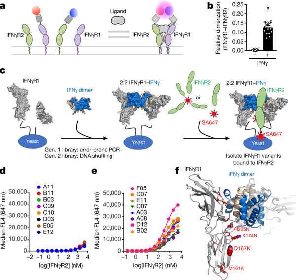
a, Cell-surface labelling of IFNγR1 (purple) and IFNγR2 (green) using Rho-11 (red circle) and DY647-labelled (blue circle) anti-GFP nanobodies is used to determine receptor dimerization. b, Relative co-tracking of Rho-11–IFNγR1 and DY647–IFNγR2 in the absence and presence of ligand. Data are mean ± s.e.m.; n = 8 (−IFNγ), n = 15 (+IFNγ); n is the number of biologically independent samples. c, Experimental design for engineering higher-affinity IFNγR1 variants. IFNγR1 (grey) is displayed on yeast and, in the presence of unlabelled IFNγ dimer (blue and tan), forms the intermediate 2:2 IFNγ–IFNγR1 complex (middle), enabling detection of variants binding to either tetrameric or monomeric IFNγR2 (green; labelled with streptavidin–Alexa Fluor 647 (SA647)). d, Using this platform, a first-generation library was generated using non-biased error-prone PCR followed by DNA shuffling. e, After a single round of selection, eight clones were titrated to estimate their relative binding to IFNγR2. f, Sites of mutations on IFNγR1 F05 (see Extended Data Fig. 1e).