Fig. 1.
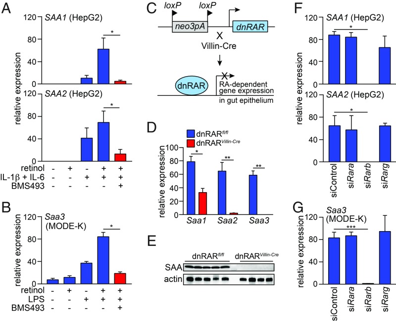
RARβ directs retinoid-dependent SAA expression. (A and B) qPCR analysis of SAA (human) and Saa (mouse) expression. (A) SAA1 and SAA2 expression in HepG 2 cells treated for 24 h with retinol, IL-1β, IL-6, and the RAR inhibitor BMS493. (B) Saa3 expression in MODE-K cells treated for 24 h with retinol, LPS, and BMS493. n = 4 replicates/group; data represent two independent experiments. (C) Selective disruption of RAR activity in intestinal epithelial cells. Knock-in mice carry a neomycin resistance gene and 3 loxP-flanked polyadenylation sequences located upstream of an ORF encoding a dominant negative (dn)RAR. Breeding to Villin-Cre mice results in selective expression of the dnRAR in intestinal epithelial cells. (D) qPCR analysis of Saa transcripts in the small intestines of dnRARfl/fl mice and dnRARVillin-Cre mice. n = 4 mice/group; data represent two independent experiments. (E) Western blot of SAA in the small intestines of dnRARfl/fl mice and dnRARVillin-Cre mice. (F and G) qPCR analysis of SAA (human) and Saa (mouse) expression after siRNA knockdown of individual Rar isoforms. (F) SAA1 and SAA2 transcripts were analyzed in HepG2 cells treated with retinol, IL-1β and IL-6 as in A, and (G) Saa3 expression was analyzed in MODE-K cells treated with retinol and LPS as in B. n = 3 replicates/group; data represent two independent experiments. Means ± SEM are plotted. *P < 0.05; **P < 0.01; ***P < 0.001 as determined by Student’s t test.

