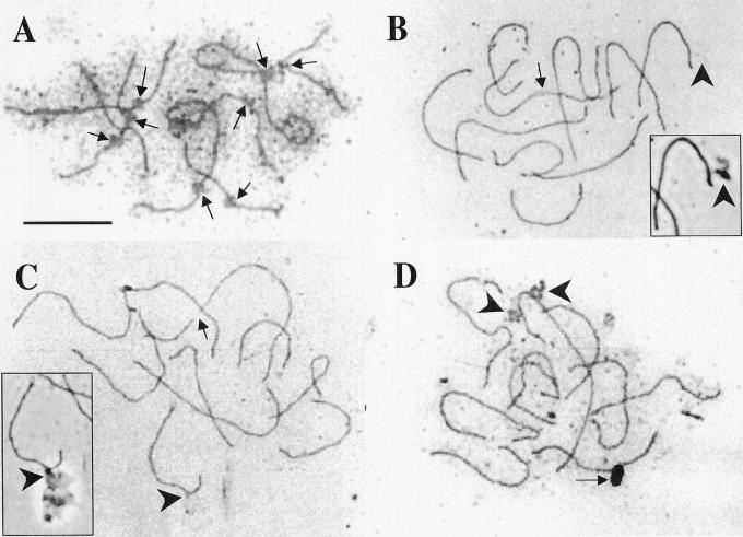
Figure 7.
SCs of SB. A through D, Silver-stained SC spreads. All photographs show bright-field images except where noted. A, Partial set of SCs in which kinetochores (arrows) are readily visible. B and C, Complete SC sets. In B, one SC contains a region of incomplete synapsis (arrow), whereas in C one SC appears to be broken into two pieces (arrow). In B and C, the sixth longest SC is associated with an amorphous structure of unknown origin (large arrowheads). These structures, most readily visualized by phase-contrast microscopy (see insets of B and C), may be part of the nuclear scaffold. In D, the longest chromosome appears to be associated with remnants of the nucleolus (large arrowheads). Although the dark oval object near the bottom of this bright-field image (arrow) looks like it could be a cellular structure, examination of this SC set by phase-contrast microscopy (photo not shown) suggests that the object is a small piece of glass. Scale bar = 10 μm.

