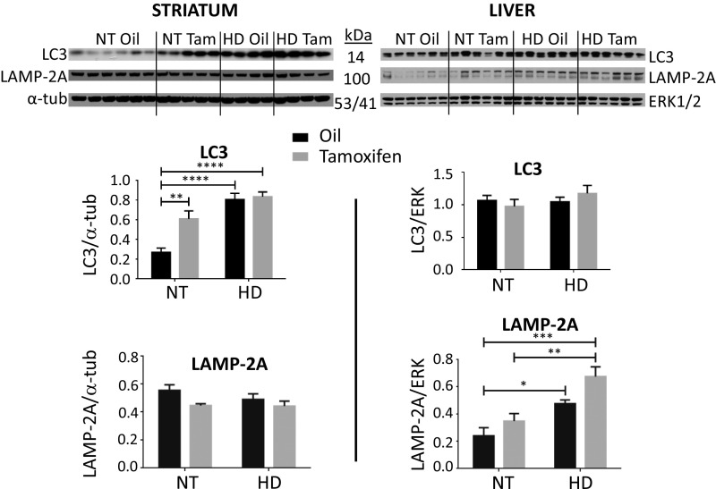
Fig. 4.
IKKβ knockout and mutant HTT exon 1 protein expression impact levels of autophagy proteins in vivo. R6/1 (HD) or NT WT control male mice containing the tamoxifen-inducible Cre and floxed alleles of IKKβ were treated with tamoxifen or oil vehicle control for 1 wk starting at 10 wk to knock out IKKβ in striatum and liver. At the termination of the study at 16 wk, Western analysis of NT and HD soluble fractions was used to examine levels of autophagy proteins LC3 and LAMP-2A relative to loading controls α-tubulin (striatum) or ERK1/2 (liver). LC3 I was detected in striatum, while LC3 I and II were observed in liver. LC3 I was quantitated for both tissues and was found in striatum, but not in liver, to be significantly increased with IKKβ knockout or with transgene expression. LAMP-2A levels were unchanged in striatum but were significantly increased in HD mouse liver. Western images, shown, were quantitated; *P < 0.05, **P < 0.01, ***P < 0.001, ****P < 0.0001 values represent means ± SEM. Statistical significance was determined by one-way ANOVA with Bonferroni posttesting.

