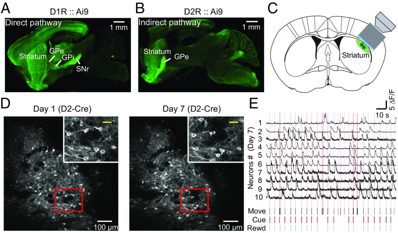
Fig. 2.
Chronic recording of activity of the same populations of D1R and D2R neurons during learning of a cued motor task. (A) Sagittal brain section obtained from a D1-Cre mouse that was crossed with the Ai9 tdTomato Cre reporter line, showing that tdTomato-labeled D1R SPNs in the direct pathway selectively send projections to the GPi and SNr. (Scale bar, 1 mm.) (B) Sagittal brain section from a D2-Cre mouse that was crossed with the Ai9 tdTomato Cre reporter line, showing that D2R SPNs of the indirect pathway specifically project to the GPe. (Scale bar, 1 mm.) (C) Schematic diagram showing the location of the objective lens and cannula for chronic imaging of DLS neurons. (D) Example fluorescence images of DLS neurons expressing GCaMP6s in D2R neurons on day 1 and day 7 (last day) of training. (Scale bars, 100 μm.) (Insets) Magnification of area in red box. (Scale bars, 30 μm.) (E) Traces of relative Ca2+ transients (ΔF/F) for 10 sample neurons and simultaneous recordings of behavioral events on day 7 in an example D2-Cre mouse expressing GCaMP6s. Pink lines represent the lever movement zone. Black, lever movement (Move); red, Cue; blue, reward (Rewd). (Horizontal bar, 10 s; vertical bar, 5 ΔF/F).

