Fig. 4.
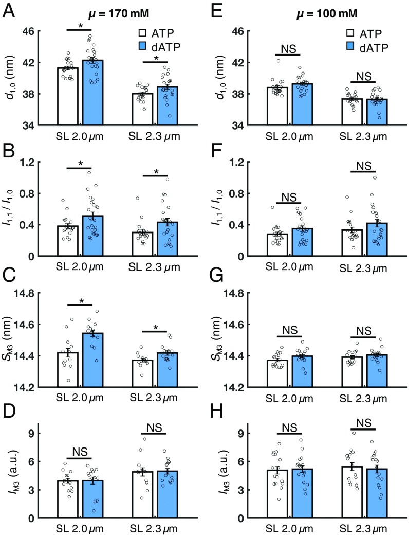
Effects of dATP on lattice geometry and myofilament structure in resting cardiac muscle (pCa 9.0). At physiological ionic strength (μ = 170 mM), dATP (blue bars) significantly increases the lattice spacing (A, d1,0), the intensity ratio of the primary equatorial reflections (B, I1,1/I1,0), and the spacing of the M3 reflection (C, SM3) at both short and long SLs compared with ATP (white bars), but does not affect the intensity of the M3 reflection (D, IM3) at either SL. However, at low ionic strength (μ = 100 mM), these effects are diminished at both short and long SLs (E–G) and the IM3 remains unaffected by dATP (H). Error bars represent SEM for n ≥ 14 preparations. *P < 0.05. a.u., arbitrary units; NS, not significant. (Numerical values are reported in SI Appendix, Table S1.)

