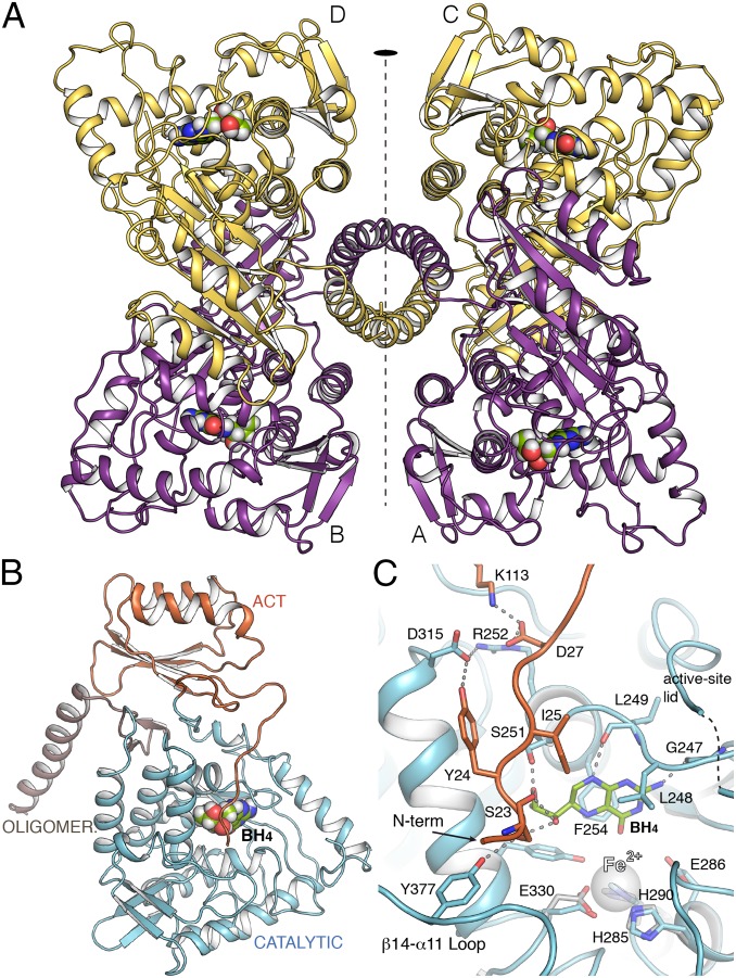
Fig. 1.
Crystal structure of human PAH. (A) Ribbon representation of hPAH in complex with BH4 at the four active sites (Tet2). The tetramer is formed as a dimer of dimers by a twofold symmetry (depicted). BH4 cofactor is drawn as spheres. (B) The hPAH monomer with each domain colored differently. (C) Detailed view of the active site of hPAH in complex with BH4 (green sticks). Relevant residues are represented as capped sticks and labeled. The FeII cation, not present in the structure, is represented as a transparent sphere for comparison purposes. Its location is obtained by direct superposition with the CD of hPAH (obtained in this work). Residues from this structure involved in the interaction with FeII cation are shown in gray sticks.

