Fig. 6.
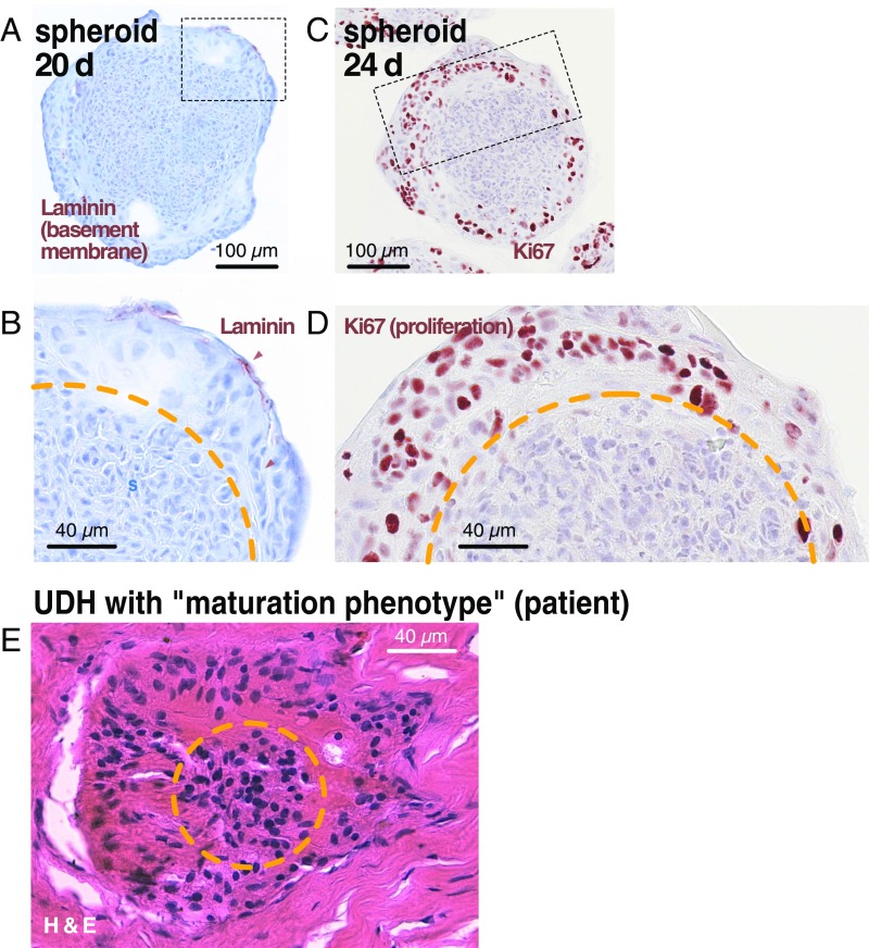
Similarities in morphology of young spheroids and UDH with “maturation” phenotype. (A–D) A 20-d-old spheroid and a 24-d-old spheroid stained for laminin and Ki67, respectively. Note the clear separation between central and peripheral zones. In the peripheral zone, cells deposit laminin at the periphery and proliferate, being surrounded by a pale, hyaline substance with indistinguishable cell borders (compare with Fig. 1B). (E) Small focus of UDH from a patient featuring a maturation phenotype. This name is based on the assumption in the literature that the peripheral, more pale nuclei surrounded by hyaline substance represent cells that undergo a maturation process and become more differentiated toward the lesion center. Our spheroid results suggest that, instead, the peripheral cells are derived from and grow out from the core. The orange line marks the border between dense central and loose peripheral zones.

