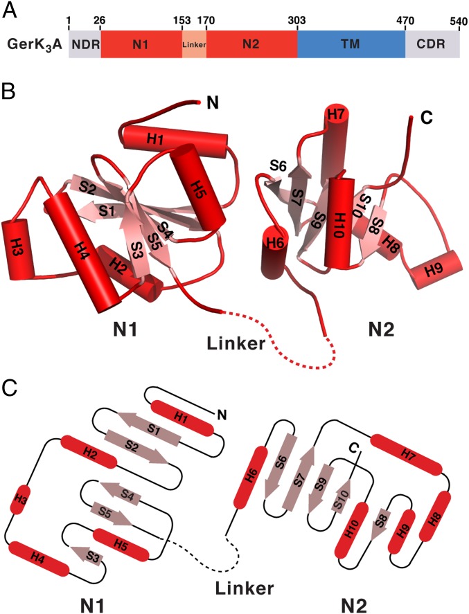
Fig. 1.
Crystal structure of GerK3ANTD. (A) Schematic representation of the domain organization of GerK3A. GerK3ANTD (residues 26–302) comprises N1, linker, and N2 domains. The boundaries of the N1 and N2 domains are defined according to results presented here. (B) Ribbon diagram of GerK3ANTD, with the secondary structure elements labeled. α-helices and β-strands are shown in red and pink, respectively. The disordered linker region is marked by a dashed line. (C) Topology diagram of GerK3ANTD. Cylinders and arrows represent α-helices and β-strands, respectively.

