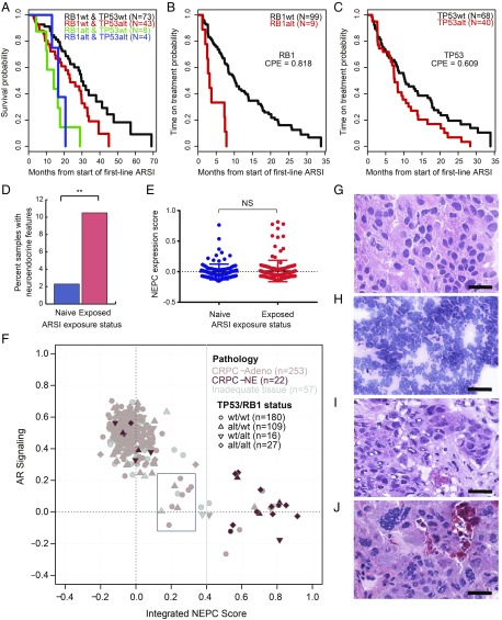
Fig. 5.
Integrative analysis incorporating histopathology, transcript-based assessment of AR signaling and NEPC score, TP53 and RB1 genomic status, and clinical outcomes. (A) Kaplan–Meier analysis showing overall survival from the start of a first-line ARSI versus genomic status for TP53 and RB1 in n = 128 patients who received a first-line ARSI and underwent tissue profiling at baseline (before or within 90 d of therapy start). (B and C) Kaplan–Meier analysis showing time on treatment with a first-line ARSI by genomic status for RB1 and TP53. P values were generated from the log-rank statistic. (D) Frequency of histopathologic neuroendocrine features in pre- versus post-ARSI samples, among patients who received an ARSI at some point during their treatment history. Patients who were not reported to have received an ARSI at any point were excluded. **P < 0.01. (E) NEPC expression score in pre- (n = 118) versus post- (n = 152) ARSI samples, as in D. NS, not significant. (F) AR and NEPC expression scores, histopathology (CRPC-Adeno, no NE features; CRPC-NE, histopathologic NE features) and TP53/RB1 genomic status (circle, wild type for both; diamond, both altered) for the 332 tumors with RNA-sequencing data. Ten cases (3%, blue box) had low AR and low NEPC expression scores. (G–J) Representative cases of CRPC-Adeno (G), CRPC-NE, small-cell type (H), CRPC-Adeno showing intermediate transcriptomic scores (I), and CRPC-Adeno showing a high NEPC score/low AR signaling score (J). Tumors represented in I and J were noted to have distinct nuclear features, including various degrees of nuclear pleomorphism, irregular nuclear membrane contours, and/or high mitotic activity. (Scale bars, 25 μm.)

