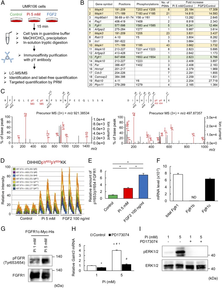
Fig. 3.
FGFR1c functions as a Pi-sensing mechanism. (A) Schematic diagram of the proteomics workflow. UMR106 cells were treated with 5 mM Pi or 100 ng/mL FGF2 for 15 min. After immunoaffinity purification with an anti-phosphotyrosine antibody, each sample was analyzed by LC-MS/MS. Three biological replicates for each sample were prepared, and an accurate amount of the proteins was measured by targeted MS. (B) The results of identification and label-free quantification of the top 20 peptides with phosphotyrosine in the order of fold increase induced by high Pi. (C) Phosphorylation of tyrosines 583 and 585 and tyrosines 653 and 654 of FGFR1 was demonstrated by the MS/MS spectrum of the m/z 921.38534 and 497.87357 ions in tryptic phophopeptides, respectively. Experiments were independently repeated two times with similar results. (D) Extracted ion chromatograms of transitions of the FGFR1 peptide with phosphorylated tyrosines 653 and 654. Each peptide in three independently prepared samples was analyzed by targeted MS using the PRM method. (E) The amount of FGFR1 peptide with phosphorylated tyrosines 653 and 654 normalized to the amount of cyclin-dependent kinase 2 (CDK2) peptide with phosphorylated tyrosine 15 in the same sample. (F) The expression level of FGFR1 subtypes evaluated by qPCR. (G) Immunoblotting with phosphorylated FGFR (tyrosine 653/654) from the lysates of UMR106 cells overexpressing Fgfr1c under 1 or 5 mM Pi for 15 min. (H) Galnt3 mRNA expression with 1 or 5 mM Pi for 48 h with or without PD173074. (I) Immunoblotting with an antibody to phosphorylated ERK1/2 from the lysates of UMR106 cells under 1 or 5 mM Pi, with or without PD173074 for 15 min. Data represent the mean ± SEM. (E) n = 3 per group; *P < 0.05 by ANOVA with a post hoc Tukey’s test. (F) n = 3 per group. ND, not detected. (G and I) Data are presented as a representative image. (H) n = 3 per group; *P < 0.05 compared with 1 mM Pi without PD173074, #P < 0.05 compared with 1 mM Pi with PD173074, †P < 0.05 compared with 5 mM Pi with PD173074 by ANOVA with a post hoc Tukey’s test.

