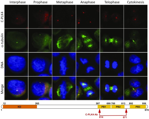
Fig. 1.
PLK4 is localized to centrosomes throughout S-phase, G2, and M-phase during the cell cycle when an antibody against C-terminal PB domains is used for immunofluorescence (IF). IF microscopy using anti-PLK4 rabbit polyclonal antibody (C-PLK4, ab137398; Abcam; to aa 614–871; red IF) and an anti–α-tubulin antibody (green IF) in synchronized cell lines permitted identification of PLK4 in centrosomes throughout the cell cycle (interphase, prophase, metaphase, anaphase, telophase, and cytokinesis). Results are illustrated for OVCAR3 ovarian cancer cells photographed at 400× original magnification. (Lower) Approximate location of the commercially available (C-PLK4) antibody used for IF above. Please note differences in PLK4 distribution compared with Fig. 2 based on phosphorylation status and epitopes recognized.

