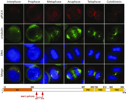
Fig. 2.
Localization of phospho-PLK4 to centrosomes, kinetochores, and midbody varies with the phase of the cell cycle when an antibody to the N-terminal PEST domain is used. Phospho-serine305-PLK4 is identified first in centrosomes of interphase and prophase cells followed by kinetochores (metaphase and anaphase) and, subsequently, the cleavage furrow (telophase) and midbody (cytokinesis) of synchronized cells by immunofluorescence (IF) microscopy using an anti–phospho-serine305-PLK4 rabbit polyclonal antibody (our Ab#3 to a PLK4-serine305 peptide; red IF). Anti–α-tubulin antibody (green IF) identifies centrosomes, spindle apparatus, and midbody. DAPI stain (blue) identifies DNA. Results are illustrated for OVCAR3 ovarian cancer cells photographed at 400× original magnification. (Lower) Schematic diagram illustrates approximate location (arrows) of our rabbit polyclonal antibody (Ab#3, anti-phospho-PLK4) for phospho-PLK4. Similar results were obtained with a second, independently produced anti–phospho-serine305-PLK4 antibody (14299) from the laboratory of G.P.N.

