
Figure 1.
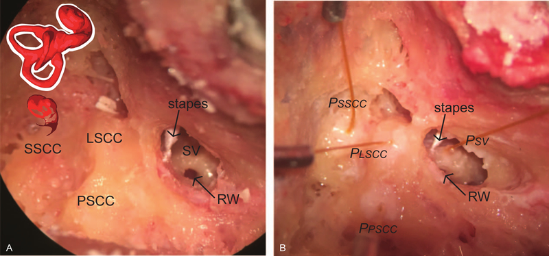
Photomicrographs of the specimen preparation. Temporal bones of intact cephali underwent mastoidectomy with wide facial recess. Scala vestibuli near the oval window (SV), lateral semicircular canal (LSCC), posterior semicircular canal (PSCC) and superior semicircular canal (SSCC) were blue lined in preparation for the intracochlear pressure probes (A). Pressure probes (PSV, PLSCC, PPSCC, PSSCC) were placed in cochleostomies in SV and canals, retro-reflective glass beads placed on the stapes for LDV measurements (B).