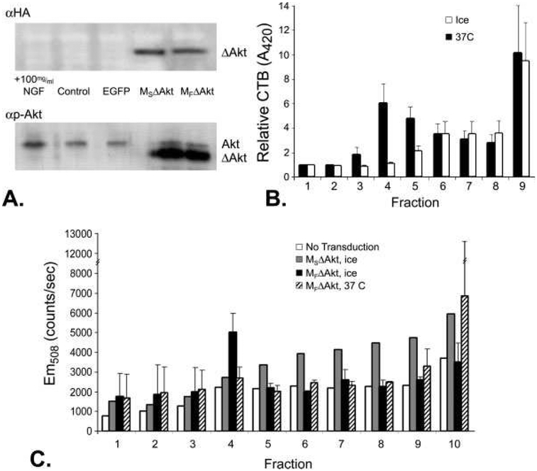
Fig. 1.
DRG neurons transduced by p.MSΔAkt and p.MFΔAkt display different subcellular targeting of the Akt/EGFP fusion protein. A: Western blot detection of HA-epitope tag or phospho-AktS473 (p-Akt) of neuronal cultures transduced with MSΔAkt or MFΔAkt revealed efficient and similar levels of expression of the myristoylated transgene (ΔAkt). Addition of NGF (positive control) increased the levels of p-Akt, whereas cultures transduced with p.EGFP had levels of p-Akt similar levels in to control cultures. B: Peroxidase activity from CTB-HRP was detected by measuring the level of oxidized ABTS, which absorbs light at 420 nm. Neuronal cultures lysed at 0°C had high levels of peroxidase activity in fraction 4, indicating the presence of buoyant lipid rafts. Incubation of neuronal cultures in Triton X-100 at 37°C, known to disrupt lipid rafts, abolishes the peak in peroxidase activity in fraction 4. C: Detection of EGFP (Em508) from sucrose gradient fractions indicated lipid raft accumulation of MFΔAkt. In cultures incubated on ice, which maintains the integrity of the lipid rafts, greater amounts of EGFP in the lipid raft fraction (fraction 4) of neuronal cultures that expressed the MFΔAkt fusion protein were detected compared with cultures that expressed the MSΔAkt fusion protein. Disruption of lipid rafts with Triton X-100 at 37°C before fractionation decreased the amount of Akt/EGFP fusion protein in fraction 4 of neurons that expressed MFΔAkt fusion protein to control levels.

