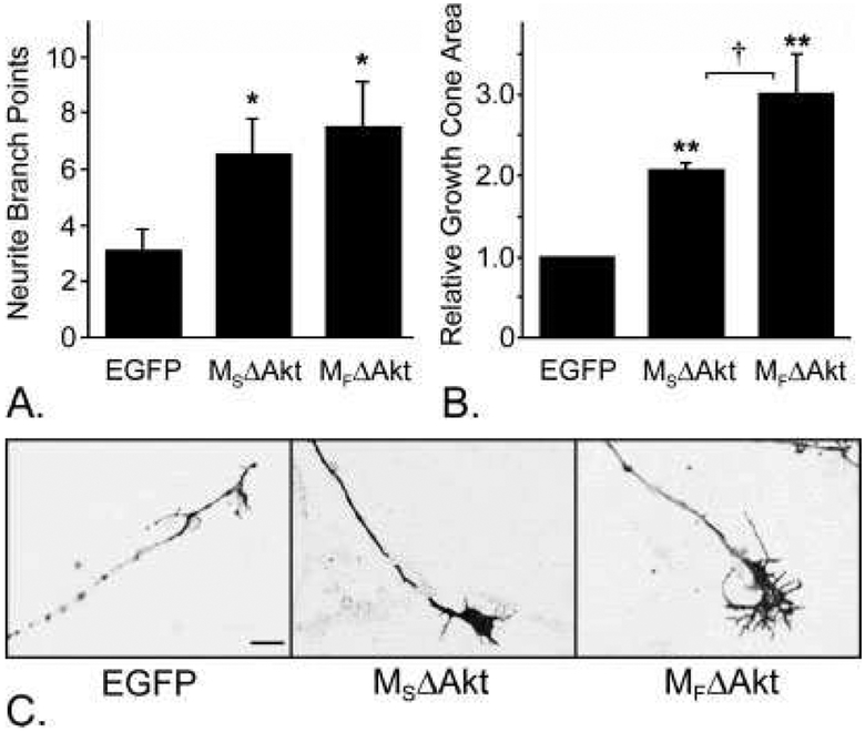
Fig. 2.
Expression of myristoylated Akt promoted axonal branching and growth cone expansion. A: DRG neurons that expressed MSΔAkt (n = 14) or MFΔAkt (n = 20) had significantly more axonal branch points than neurons that expressed the control protein, EGFP (n = 32; P < 0.0001 for each), but there was no significant difference in axonal branching between the two Akt constructs (P = 0.17). B: Neurons that expressed CA-Akt throughout the plasma membrane (MSΔAkt; n = 54) had significantly larger growth cone areas than control neurons that expressed EGFP (n = 97; P = 0.007). Neurons that expressed lipid raft-targeted CA-Akt (MFΔAkt; n = 92) had significantly larger growth cones than neurons that expressed either MSΔAkt or EGFP (P = 0.045 and P = 0.005, respectively). C: Representative photomicrographs growth cones from transduced DRG neurons.
*P < 0.05, **P < 0.01 compared with EGFP, †P < 0.05 between MSΔAkt and MFΔAkt. Scale bar = 10 μm.

