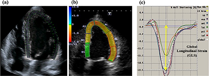
Fig 2. Example of the measurement of left ventricular (LV) global longitudinal strain (GLS) with a two-dimensional speckle-tracking echocardiography (2DSTE) in a patient with chronic total occlusion who underwent successful percutaneous coronary intervention (PCI).
Figure (a) and (b) showed apical four-chamber view. White broken lines indicated the LV global strain curves for GLS. Yellow arrow indicated the mean LV global peak longitudinal strain; GLS: −22.5% (c).

