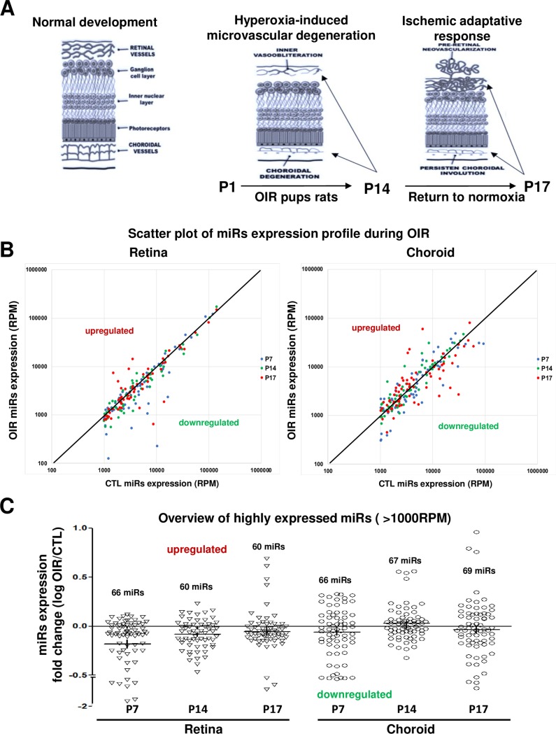
Fig 1. Overview of differential-expression profile of miRNAs in the retina and choroid of rats subjected to OIR.
A. Schematic representation of OIR-induced pathological effects on microvascular development in the retina and choroid at different postnatal ages (P). Hyperoxia exposure leads to vessel degenerations (P1 to P14). At P17 (return to normal room air for 3 days), one observes pathological neovascularization (NV) pre-retinally associated with persistent vessel degeneration in the choroid. B and C. Scatter plot of miRNA expression alteration during OIR (RPM) (B) and overview of highly expressed miRNAs (more than 1000 reads per million [RPM]) in retinal and choroidal tissues and their differential-expression profile (log fold change: OIR/CTL) (C) assessed by next generation sequencing (NGS). Data are log RPM fold change ratio (OIR/CTL) of 5 rats/group.

