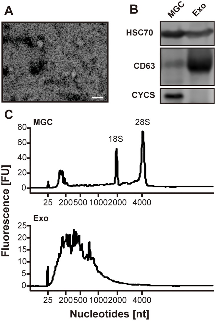
Fig 1. Detection of exosome-like vesicles in the exosomal fraction isolated from pFF.

(A) Representative photograph of vesicles in the exosomal fraction isolated from pFF observed using transmission electron microscopy. The scale bar indicates 100 nm. (B) Western blotting analysis for HSC70, CD63, and CYCS. MGC, mural granulosa cell; Exo, exosomal fraction. (C) Representative electropherograms observed using bioanalyzer. FU, fluorescence intensity units; nt, nucleotides.
