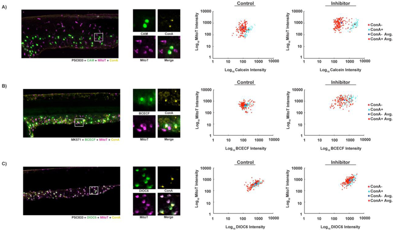
Figure 6. Calcein and BCECF accumulate in HR cells, and DiOC6 accumulates in MR cells.
Confocal projections of embryos exposed to ABC transporter substrate (green), general ionocyte marker mitoT (magenta), and HR cell marker conA (yellow). Graphs indicate fluorescence intensity of epidermal cells in arbitrary fluorescence units. (A) Calcein accumulates to higher levels in mitoT+/conA+ than mitoT+/conA- cells in both absence (p < 0.0001, n = 251 cells from 3 embryos) and presence of PSC833 (p < 0.0001, n = 264 cells from 3 embryos). (B) BCECF accumulates in mitoT+/conA+ cells and some but not all mitoT+/conA- cells. BCECF intensity is significantly different in conA- and conA+ cells in the absence (p = 0.0025, n = 238 cells from 3 embryos) and presence of MK571 (p < 0.0001; n = 214 cells from 3 embryos). (C) DiOC6 accumulates in all mitoT+ cells and is significantly different in conA- and conA+ cells in the absence (p = 0.0076, n = 246 cells from 3 embryos) and presence of PSC833 (p < 0.0001; n = 258 cells from 3 embryos).

