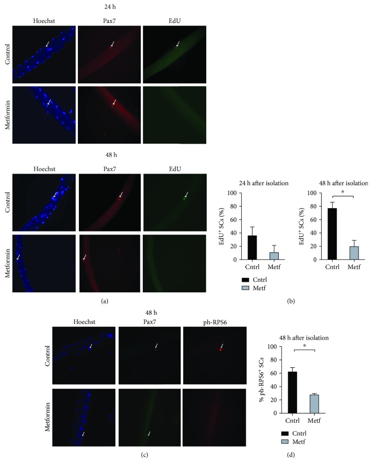
Figure 7.
Metformin delays SC activation upon administration in vivo. (a) Single myofibers were isolated from control and metformin-treated C57BL/6 mice and cultured in vitro in Tyrode's medium containing an EdU labeling agent. The SCs associated with the myofibers were analyzed by immunofluorescence microscopy for the expression of Pax7 and the incorporation of EdU after 24 h and 48 h of culture. (b) Percentage of EdU-positive SCs associated with single myofibers isolated from control and metformin-treated C57BL/6 mice. The SCs associated with the myofibers were analyzed by immunofluorescence microscopy for the expression of Pax7 and the incorporation of EdU after 24 h and 48 h of culture (n24h cntrl = 48 SCs, n24h metf = 40 SCs, n48h cntrl = 40 SCs, and n48h metf = 36 SCs). (c) Single myofibers were isolated from control and metformin-treated C57BL/6 mice and cultured in vitro. The SCs associated with the myofibers were analyzed by immunofluorescence microscopy for the expression of Pax7 and ph-RPS6 after 48 h of culture. (d) Quantitation of the percentage of myofibers associated SCs positive for ph-RPS6 after 48 h in culture. Myofibers were isolated from control and metformin-conditioned C57BL/6 mice (n48h cntrl = 42 SCs, n48h metf = 40 SCs).

