Figure 3.
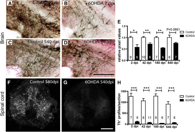
TH+ axons are inefficiently regenerated. A–D, Immunohistochemical detection of TH+ axons (black on red counterstain) in sagittal sections through a terminal field of TH+ axons ventral to population 5/6 is shown. A–D, Compared with controls (A), density of these axons is reduced at 2 dpi (B), and is more similar to age-matched controls (C) at 540 dpi (D). E, Semiquantitative assessment of labeling intensity in the area depicted in A–E indicates significant loss of innervation at all time points except the latest, 540 dpi. F, G, Spinal cross sections are shown. Compared with age-matched controls (F), immunofluorescence for TH is very low at 540 dpi (G). H, Quantification of spinal TH+ axons indicates a lack of regeneration of the spinal projection. Student's t tests (with Welch's correction for heteroscedastic data) or Mann–Whitney U tests were used for pairwise comparisons as appropriate: *p < 0.05; **p < 0.01; ***p < 0.001. Error bars represent S.E.M. Scale bars: (in D) A–D, 100 μm; (in G) F, G, 100 μm.

