Figure 4.
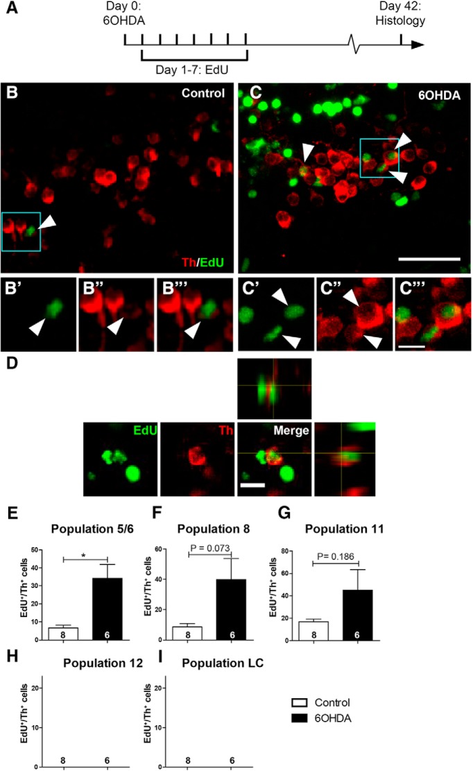
Generation of new TH+ cells is enhanced by prior ablation only in dopaminergic populations showing constitutive neurogenesis. A, The experimental timeline is given. B, C, In sagittal sections of population 5/6 (rostral left; dorsal up), EdU and TH double-labeled cells can be detected. Boxed areas are shown in higher magnifications in B′ to C‴, indicating cells with an EdU-labeled nucleus, which is surrounded by a TH+ cytoplasm (arrowheads). D, High-magnification and orthogonal views of an EdU+/TH+ cell after 6-OHDA treatment are shown. E–I, Quantifications indicate the presence of newly generated TH+ cells in specific dopaminergic brain nuclei (E–G). After 6-OHDA treatment, a statistically significant increase in the number of these cells was observed for population 5/6. Note that population 12 and LC showed no constitutive or ablation-induced EdU-labeled TH+ cells (H, I). Student's t tests with Welch's corrections, *p < 0.05. Error bars represent S.E.M. Scale bars: (in C) A, B, 20 μm; (in C‴) B′ to C‴, 5 μm; D, 10 μm.

