Figure 1.
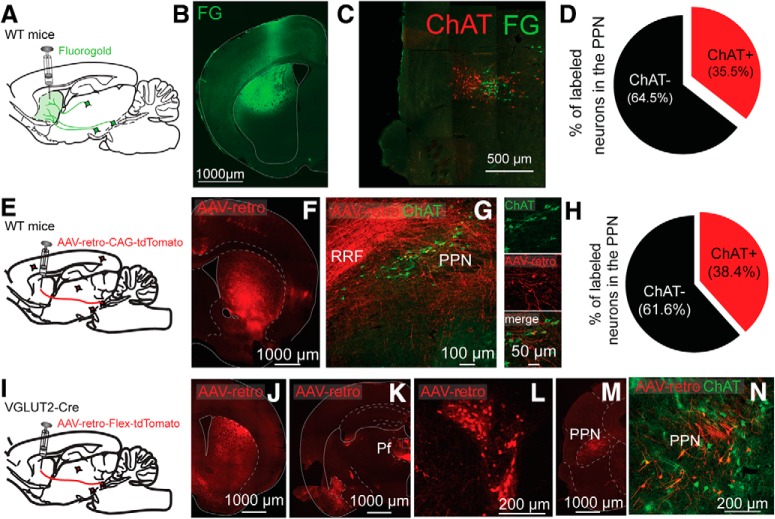
Striatal retrograde tracing reveals cholinergic and glutamatergic projections originating in the PPN. A–D, Injection of fluorogold into the striatum reveals ChAT+ and ChAT− neurons in the PPN. A, Experimental design. B, Injection site in striatum. C, Retrogradely labeled neurons in PPN delimited by ChAT immunostaining (red). D, Quantification of ChAT+/ChAT− retrogradely labeled neurons. E–H, Striatal injection of AAV retro reveals similar proportions of ChAT+ and ChAT− neurons in the PPN. E, Experimental design. F, Striatal injection site. G, Retrogradely labeled neurons in the PPN (red) combined with ChAT immunocytochemistry (green). H, Percentage of retrogradely labeled neurons in the PPN expressing ChAT. Similar numbers of retrogradely labeled ChAT+ and ChAT− neurons were found using fluorogold (D) or AAV-retro (H). I, Cre-dependent AAV-retro-td-tomato virus was injected in the striatum of VGLUT2-Cre mice. J, Striatal injection site. K–L, Retrogradely labeled neurons in the parafascicular nucleus of the thalamus and the PPN (M, N), as delimited by ChAT immunostaining (N).

