Figure 7.
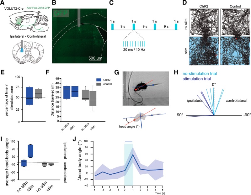
Stimulation of PPN glutamatergic axons in the striatum induces ipsilateral head movements. A, Injection of AAV2-Flex-ChR2-GFP into the PPN of VGLUT2-Cre or WT mice. B, Location of the optic fiber in the dorsal striatum. C, Schematic of stimulation parameters. D, Representative open field tracking of VGLUT2-Cre mice transduced with ChR2 in the PPN (left), compared to a control mouse (right). Top, No stimulation (“no stim”). Bottom, Laser stimulation (“stim”). E, Percentage of time spent in the stimulated compartment. Blue represents stimulation compartment. Gray represents no stimulation compartment. F, Distance traveled by the animals with or without stimulation of the PPN or controls (gray). Stimulation of PPN does not change the distance traveled by the animals in the open field (30 min session). G, Representation of the head-body (red) and tail-body (blue) axes. H, Variation (δ) of angular velocity in the nonstimulated trials (light blue) versus the stimulated trials (dark blue). I, Angular velocity in nonstimulated (no stim) or stimulated (stim) trials. J, Change of the angular velocity over time.

