
Fig. 2. AdOPG transduction changes RANKL/OPG homeostasis in primary hMSCs differentiated on Col-GAG and MC-GAG.
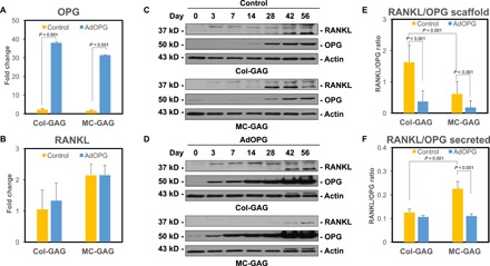
qRT-PCR of control or AdOPG-transduced primary hMSCs cultured on Col-GAG or MC-GAG materials for 14 days in osteogenic differentiation medium for (A) OPG and (B) RANKL (n = 3). Western blot of (C) control or (D) AdOPG-transduced primary hMSCs cultured on Col-GAG or MC-GAG materials for 56 days in osteogenic differentiation medium for RANKL, OPG, and β-actin. Average RANKL/OPG protein expression ratio based on (E) densitometric analysis of RANKL and OPG Western blot bands or (F) ELISA of culture supernatants over 8 weeks. Mean values are shown in bars, with error bars representing SD. Significant post hoc comparisons following analysis of variance (ANOVA) indicated with P values.