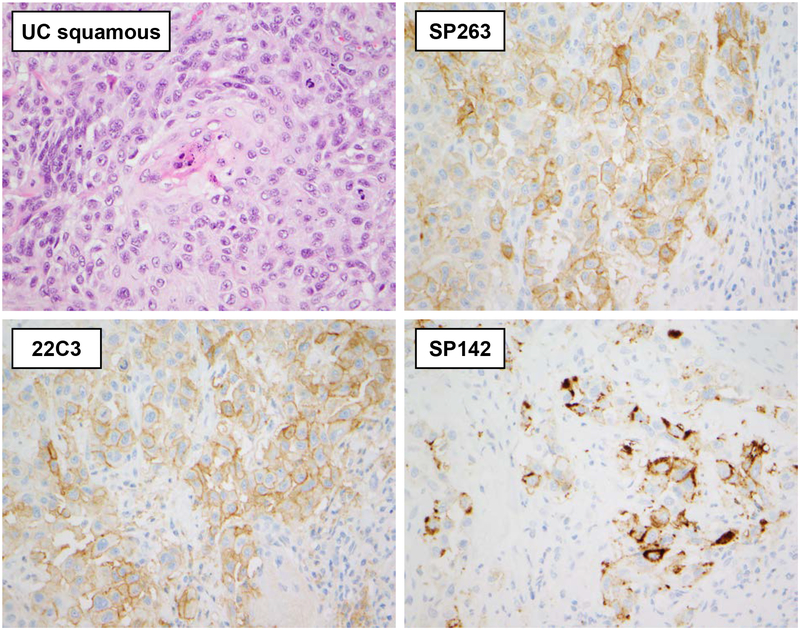
Figure 2. Staining characteristics of three PD-L1 clones in UC with squamous differentiation.
In this example of UC with squamous differentiation, membranous and focally circumferential staining of the SP263 and 22C3 PD-L1 clones in TC is evident. Clone SP142 shows a coarser and more granular TC immunoreactivity which in some cases was difficult to discriminate from IC reactivity. All 400×

