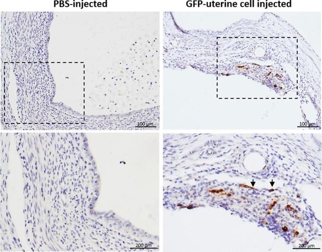
Figure 2.
Localization of uterine cells integrated into the endometriotic lesion. Representative immunohistochemical section of an endometriotic lesion demonstrating brown-stained GFP+ uterine cells found in the lesion stroma as well as integrated into blood vessels wall in the lesion (arrowheads) (n = 8, right). Representative section of a lesion from PBS-injected mice (n = 4, left). Lower panel: higher magnification of boxed areas.

