Figure 4.
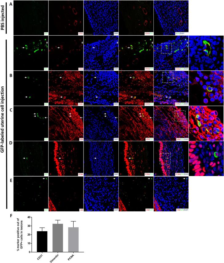
Profile of uterine cells incorporated into growing mouse endometriosis lesions. Immunofluorescent micrographs of endometriosis lesions 4 weeks post EMI. Representative sections from control (n = 4) and experimental (n = 8) lesions from different mice are shown. Co-staining of GFP-positive uterine cells integrated into a growing endometriosis lesion (green) with (A) CD31, (B) CD45 (section depicting an inflammatory cluster within the lesion), (C) vimentin, (D) PCNA, and (E) CK8 (red). Sections were counterstained with DAPI showing nuclei (blue). Arrowheads point to GFP+ integrated uterine cells. Right column: higher magnification images of dashed areas. Scale bar = 50 μm. (F) Quantification of the percentage of cells double-positive for GFP and CD31 (n = 7 lesions), vimentin (n = 7 lesions), or PCNA (n = 7 lesions).

