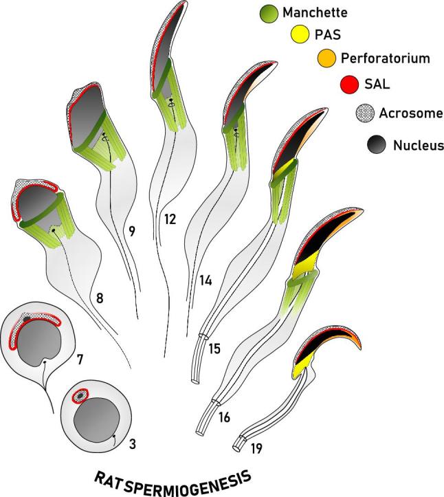
Figure 2.

Diagrammatic representation of the formation of the perinuclear theca during rat spermiogenesis. This diagram represents our current working model of the biogenesis of the perinuclear theca (PT) in falciform spermatozoa. Spermatids at representative steps of rat spermiogenesis are depicted in sagittal sections, with some structures drawn to give an appreciation of their three-dimensional range about the nucleus (i.e. manchette, PAS). The manchette (green) emerges at the onset of spermatid elongation and vanishes as the spermatid completes nuclear elongation and condensation, represented by a progressive darkening of the nucleus (gray/black). The spatial and temporal assembly of the three regions of the PT of rat spermatozoa are shown, with the subacrosomal layer (SAL, red) forming early in spermiogenesis concomitant with the acrosome (cross-hatched), and the postacrosomal sheath (PAS, yellow) appearing later as the microtubular manchette migrates distally. Note that the PAS is superimposed on the sagittal drawing to give an appreciation of its range about the caudal aspect of the spermatid nucleus following manchette descent. Similar to PAS formation, the perforatorium (orange) of the PT is also proposed to make use of the manchette for the transport of proteins into this apical compartment. Adapted from Oko [5].
