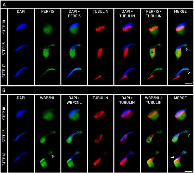
Figure 6.
Comparative developmental expression of PERF15 (A) and WBP2NL (B) with α-tubulin in spermatids throughout rat spermiogenesis using indirect immunofluorescence. Columns represent the type of analysis performed, with DAPI (blue) indicative of nuclear staining, α-tubulin (red) representative of manchette microtubules, and PERF15 (A, green) or WBP2NL (B, green) showing their respective expression. In both PERF15 and WBP2NL panels, labeling is seen in the distal cytoplasmic lobe and co-localized with α-tubulin on the manchette (*). Note that PERF15 only localizes to the perforatorial region ( ), while WBP2NL localizes to both the perforatorium (
 ) and PAS (
 ). Bars, 10 μm.

