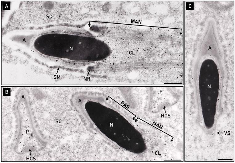
Figure 7.
Electron micrographs of rat spermatids immunolabeled with anti-PERF15 antibody. (A) PERF15 is predominantly localized to the distal cytoplasmic lobe (CL) and microtubular manchette (MAN) of a step 13 rat spermatid. (B) Accumulation of PERF15 in a pre-condensed, electron-lucent perforatorium (P), but distinctly void in the forming postacrosomal sheath (PAS) below the acrosome (A) of a step 15–16 spermatid. Labeling also occurs over the distally migrating manchette. Note the PAS is not present prior to manchette descent (see Figure 7A), but appears only after the manchette has migrated down the spermatid nucleus (N). (C) PERF15 labeling in the area of the ventral spur (VS) of a step 16 spermatid. HCS, displaced head cap segment; NR, nuclear ring; SM, Sertoli cell mantel; SC, Sertoli cell cytoplasm. Bars, 0.2 μm.

