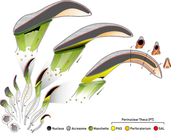
Figure 8.
Diagrammatic summary of the developmental steps proposed in the formation of the perforatorium and postacrosomal sheath (PAS) of the perinuclear theca (PT) of rat spermatozoa. Spermatids progressing through spermiogenesis are shown in sagittal sections, although some structures have been superimposed to give an appreciation of their three-dimensional architecture. Attention is paid to spermatids of step 14–16 to demonstrate the proposed assembly of the perforatorium and PAS of falciform spermatozoa. Where appropriate, representative cross-sectional views of the spermatid head provide an alternative perspective of PT biogenesis. Perforatorial-bound proteins (orange) synthesized in the distal cytoplasmic lobe of elongating spermatids migrate upwards on the manchette and pass ventrally along the spermatid head into the perforatorium. The ventral void in acrosomal material, seen in cross-sectional views, permits the passage of proteins, leading to the progressive infiltration of material in the perforatorium. Similarly, PAS-bound proteins (yellow) also make use of the manchette for the storage and transport of proteins prior to their deposition in the PAS upon manchette descent. The manchette, PAS, and acrosome are drawn to give an appreciation of their three-dimensional range in the spermatid head. Note that the subacrosomal layer (SAL, red) forms early in spermiogenesis, synchronously with the acrosome and remains adherent to the inner acrosomal membrane as the sperm head markedly expands in the creation of the perforatorium. Adapted from Lalli and Clermont [43].

