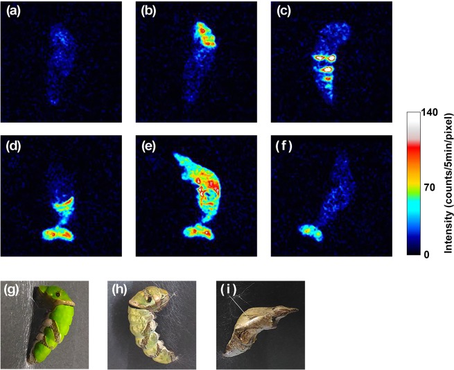
Figure 2.
Biophoton images of Papilio protenor obtained at specific time points: (a) 8 hr before ecdysis, (b) 6 hr before ecdysis, (c) 5–10 min before ecdysis, (d) 20–25 min after ecdysis, (e) 75–80 min after ecdysis, and (f) 5 hr after ecdysis. (g) Picture of the subject in the larval stage taken before biophoton measurement. (h) Picture of the pre-pupa taken directly before ecdysis (different subject, not used for biophoton imaging). (i) Picture of the subject in the pupal stage taken after biophoton measurement. Time points of (a)–(f) are designated in Fig. 1(b) with corresponding characters and arrows.

