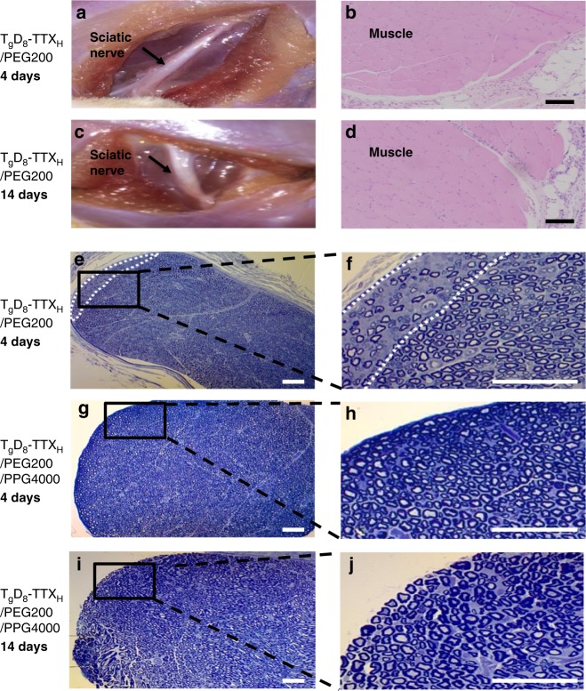
Fig. 7.
Tissue reaction to materials. a, c Representative photographs of the site of injection upon dissection 4 and 14 days after injection of 25 mg of TgD8–TTXH in 0.5 mL of PEG200. b, d Representative hematoxylin–eosin stained sections of muscles and adjacent loose connective tissue 4 and 14 days after injection of 25 mg of TgD8–TTXH in 0.5 mL of PEG200. Scale bars, 100 µm. e–j Toluidine blue stained sections of nerve after injection of formulated TgD8–TTXH. Scale bars, 50 µm. e, f Minimal peripheral injury (area enclosed by white dotted lines) seen in 1 of 3 nerves 4 days after injection of 25 mg of TgD8–TTXH in 0.5 mL of PEG200. g–j Representative toluidine blue stained sections of nerve 4 days (g, h) and 14 days (i, j) after injection of 25 mg of TgD8–TTXH in 0.5 mL mixture of PEG200 and PPG4000 (5/95, v/v), showing no injury. Data are representative of 3 animals in each group

