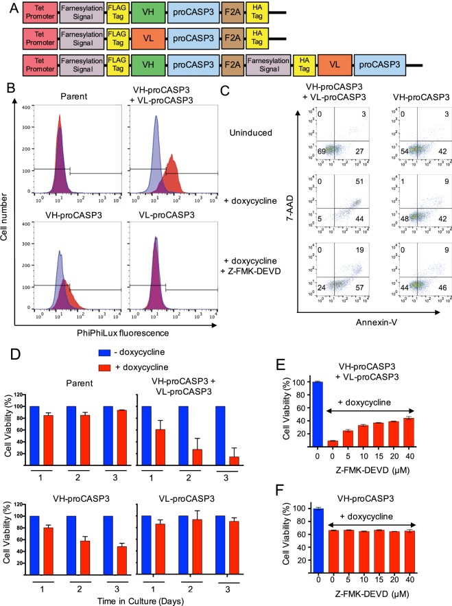
Figure 1.
Activation of apoptosis by anti-RAS iDAb-procaspase3 fusion proteins. A schematic diagram of the AIDA expression plasmids (using the tetracycline-inducible pUHD10 plasmid) controlled by the tetracycline operator (tet-promoter) is shown in panel (A). Each expressed protein has an amino terminal farnesylation signal followed by a FLAG-tag and the iDAb (VH or VL) fused to pro-caspase-3 followed by a viral F2A sequence and a HA-tag. In the bottom line, the protein is a fusion of VH-proCASP3 and VL-proCASP3 (VH-proCASP3 + VL-proCASP3). Mutant clones were also made with mutant VH and mutant caspase-3. The activation of caspase-3 was assayed using PhiPhiLux fluorogenic substrate (panel B). Clones of each type, including the parent HT1080TetOn line, were cultured in doxycycline for 48 hours and incubated with substrate and analyzed by flow cytometry. Blue = cells without doxycycline; red = cells with doxycycline. In panel C, cells carrying VH-proCASP3 + VL-proCASP3 or VH-proCASP3 were treated with and without doxycycline, and with both doxycycline plus caspase-3 inhibitor (Z-FMK-DEVD, at 40 μM) for 48 hours and apoptotic status compared by flow cytometry with Annexin-V and 7-AAD. The numbers refer to cell numbers in each indicated quadrants. Panel D shows averaged viability analysis of three clones from each stable cell type (clones A–C) with or without doxycycline induction measured over three days using the Prestoblue (Invitrogen) resazurin-based reagent. Red bars are with and blue bars are without doxycycline. The dose response to the inhibitor, Z-FMK-DEVD, was measured over a range of concentrations to 40 μM with cells grown for 48 hours, in the presence or absence of doxycycline and with and without inhibitor. VH-proCASP3 + VL-proCASP3 cells (panel E) and VH-proCASP3 only cells (panel F). Error bars in graphs shown in panels (D–F) represent standard deviation from the mean.

