Fig. 1.
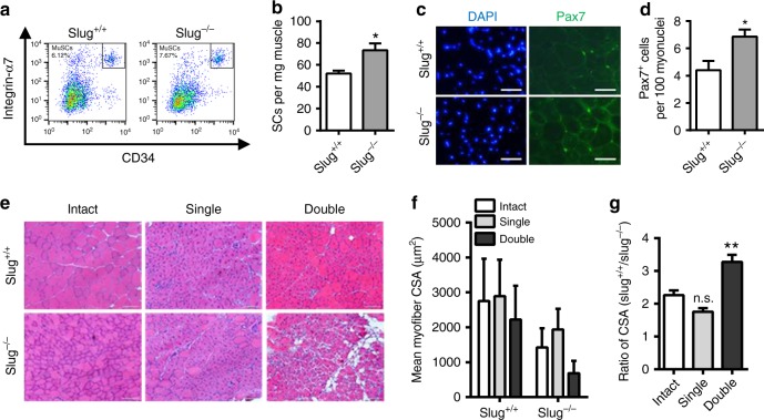
Slug deficiency repairs regenerative capacity of SCs during serial muscle damage. a Representative flow cytometric analysis of the frequency of SCs (CD45−/CD11b−/CD31−/Sca1−/Integrin-α7+/CD34+) subpopulation in Slug+/+ and Slug−/− mice. b Yield of SCs per milligram (mg) of muscle from Slug+/+ and Slug−/− mice (n = 6 mice per group). *p < 0.05 by student’s t-test. c Representative IHC images of Pax7+ SCs within the intact tibialis anterior (TA) muscles of Slug+/+ and Slug−/− mice. DAPI was used as nuclear counterstaining. Scale bar, 100 μm. d Quantification of Pax7+ SC numbers in c. *p < 0.05 by student’s t-test. e H&E staining of the intact and injured TA muscles in Slug+/+ and Slug−/− mice (n = 5 mice per group). For single injury, TA muscles were harvested at day 10 after BaCl2 injection. For double injury, mice were recovered for 1 month after the first BaCl2 injection at TA muscles. A second BaCl2 injection was administered thereafter. TA muscles were harvested 10 days after the second BaCl2 injection. Scale bar, 100 μm. f Quantification of the mean myofiber cross-sectional area (CSA, μm2) of the intact and BaCl2-injured TA muscles of Slug+/+ and Slug−/− mice. g Ratio of myofiber CSA in the intact and BaCl2-injured TA muscles between Slug+/+ and Slug−/− mice. **p < 0.01 by student’s t-test (n.s., not significant). All these experiments were independently repeated three times with similar results. Data are presented as mean ± SEM. Also see Supplementary Fig. 1. Source data are provided as a Source Data file

