Figure 2.

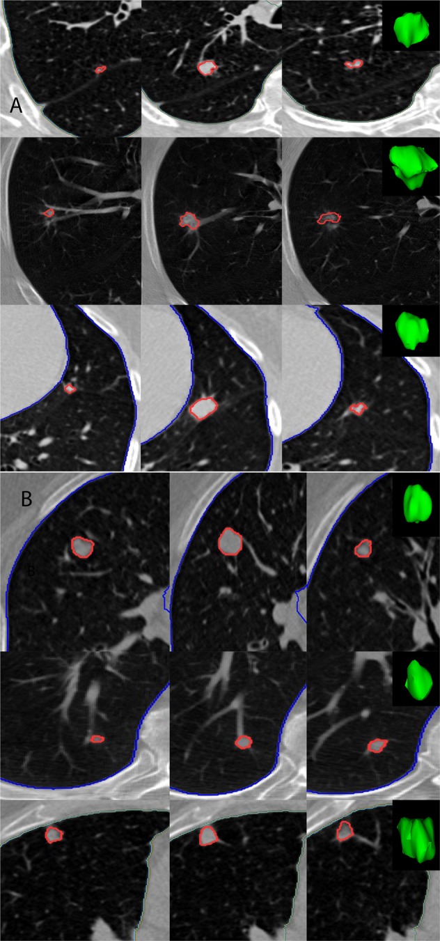
Representative 2D slices of patient LDCT with 3D rendering of the pulmonary nodule of interest, where each row corresponds to a patient (A) Patient with confirmed malignant nodule (B) Patients with benign nodules, as reported by NLST.

Representative 2D slices of patient LDCT with 3D rendering of the pulmonary nodule of interest, where each row corresponds to a patient (A) Patient with confirmed malignant nodule (B) Patients with benign nodules, as reported by NLST.