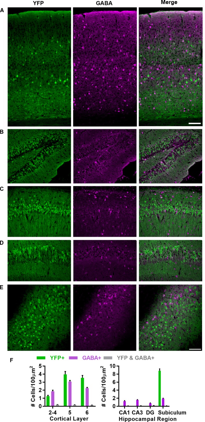
Figure 1.

Slick‐H YFP expression in cortex, hippocampus, and subiculum. (A) Slick‐H mice express Cre and a YFP reporter (green) in projection neurons of the visual cortex with limited overlap with GABA+ cells (magenta). (B–E) Slick‐H mice express Cre and a YFP reporter in projection neurons of the hippocampus with limited overlap with GABA+ cells in the dentate gyrus (DG) (B), CA1 (C), CA3 (D), and subiculum (E). (F) Quantification of overlap between YFP and GABA+ cells across cortical layers (left) and hippocampal regions (right). YFP+ cell density in DG, CA1, and CA3 not quantified due to unclear cell boundaries with high density. Scale bars (in A and E) = 100 μm.
