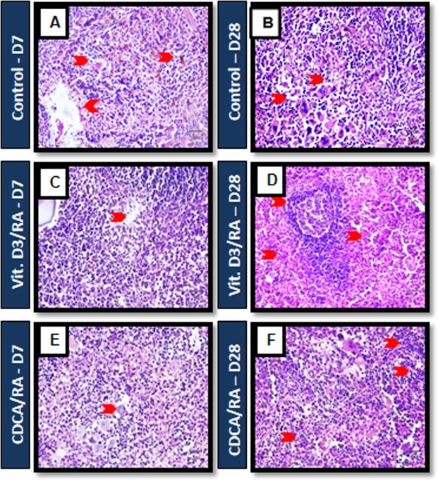
Figure 5.
Mouse infected with L. donovani splenic tissue histological staining of H and E. (A,B) Control mice Day 7 and Day 28: L. donovani infected mice showed reactive follicular hyperplasia, including expansion of marginal zone around the follicle and immature granuloma (C,E) mice treated with Vit.D3/RA and CDCA/RA displayed reactive hyperplasia of lymphoid follicles and early development of granuloma in the red pulp by day 7 of the PT. (D) 28 PT, mice treated with Vit.D3/RA showed effective response through the recruitment of red pulp with small lymphoid follicles and absence of granuloma formation. Similarly, mice treated with CDCA/RA (F) showed shrinkage in white pulp and the development of red pulp with no granuloma (arrow head, respectively), n = 6.

