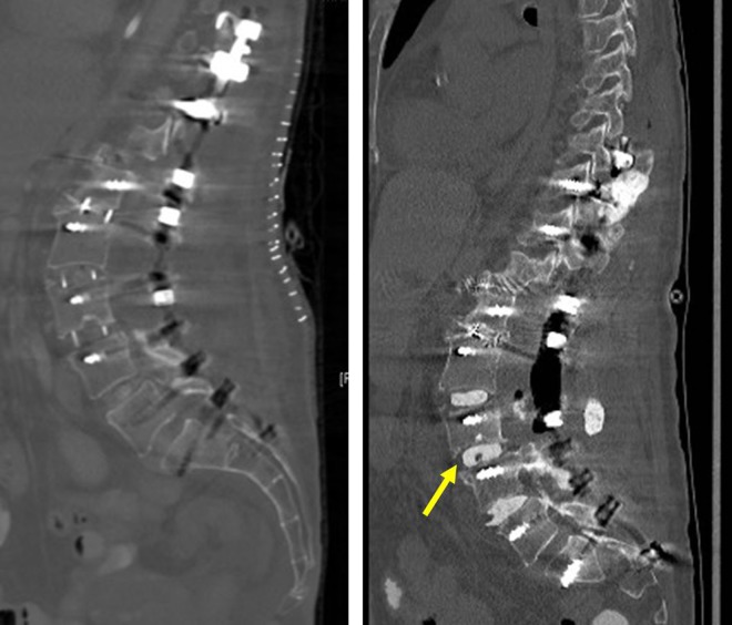
Figure 7.
Patient 3: (left) computed tomography (CT) scan sagittal image obtained after lateral surgery showing polyetheretherketone (PEEK) cages at L2-5 levels. The bowel was stuck to the L3-4 PEEK. (right) CT scan sagittal image showing removal of the PEEK cage and use of polymethylmethacrylate (PMMA) antibiotic cement spacers (yellow arrow).

