Figure 2.

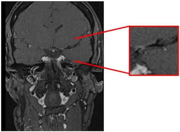
Cranial MRI, coronal T1-weighted-(dark-blood) sequence demonstrating contrast enhancement of the vessel wall in the first segment of the left middle cerebral artery.

Cranial MRI, coronal T1-weighted-(dark-blood) sequence demonstrating contrast enhancement of the vessel wall in the first segment of the left middle cerebral artery.