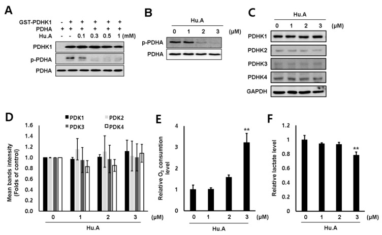
Figure 2.
Hu.A reduced the PDHK1 activity and promoted oxidative phosphorylation (OXPHOS) in DLD-1 cells. (A) In vitro PDHK1 kinase assay was performed. (B, C) DLD-1 cells were treated with Hu.A in serum-free medium for 4 h. The levels of phosphorylated pyruvate dehydrogenase E1α subunit (PDHA) (B), and PDHK1-4 (C) were analyzed using western blot assay. PDHA (B) and GAPDH (C) were used as loading controls. (D) The intensity of bands (PDHK1-4/GAPDH) from three independent experiments was measured and indicated by mean ± SD. (E) DLD-1 cells were treated with Hu.A in serum-free medium for 6 h. O2 consumption rate was measured by using commercially available Oxygen Consumption Rate Assay Kit. (F) Lactate production was measured by lactate fluorometric assay kit (right panel). The data are shown as mean ± SD, respectively. **, p < 0.01 compared with control.

