Figure 7.
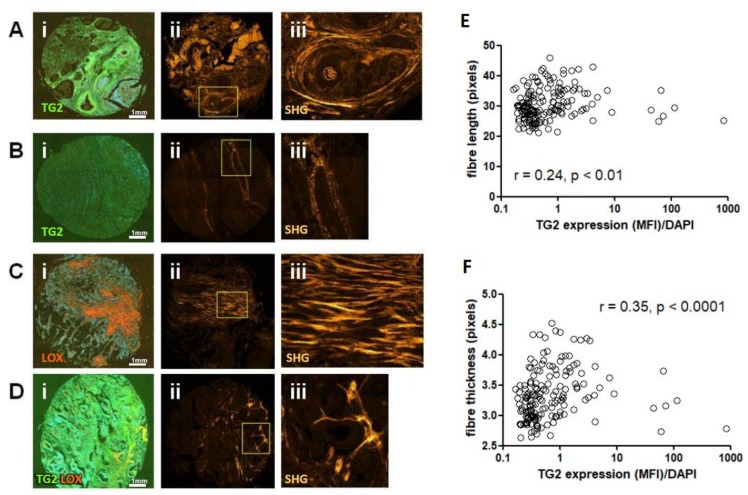
TG2, LOX and co-localisation of both correlate with collagen structural parameters determined by SHG. High TG2 expression (A, green staining in panel i) was associated with regions of thicker collagen fibres which surround the epithelium (A, panel ii and magnified image panel iii). By contrast, low TG2 expression (B, panel ii) was associated with regions of thinner fibres (panel ii and magnified image panel iii). High LOX staining (C, red staining in panel i) associated with thick fibres extending extensively throughout the stroma (panel ii and magnified image panel iii). Areas of high co-localisation of both enzymes (D, panel i, yellow colouration) showed distinctive, localised, thickened fibre bundles (D, panel ii and magnified image panel iii). Correlation analysis of staining and SHG imaging showed a significant relationship between TG2 and fibre length (E), and fibre width (F).

