Figure 2.
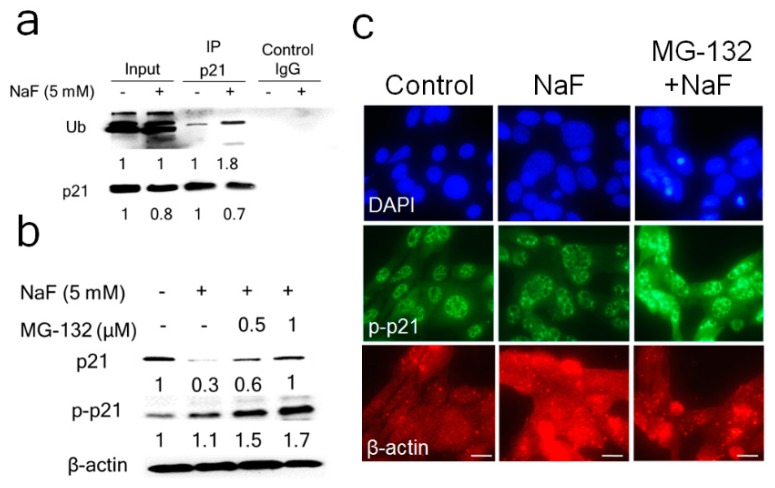
Fluoride increased Ub-p21 binding and MG-132 reversed the fluoride-mediated p21 protein decrease. (a) LS8 cells were treated with NaF (5 mM) for 6 h. Protein was immunoprecipitated using anti-p21 antibody and ubiquitinated-p21 (Ub-p21) was detected in the precipitated fraction by the anti-Ubiquitin antibody. Fluoride treatment increased Ub-p21 levels. IgG was used as the negative control. The numbers show relative protein expression vs. Controls (0 mM NaF). IP lanes were quantified separately from input lanes. (b) LS8 cells were treated with MG-132 (0.5–1.0 μM) for 2 h prior to NaF (5 mM) treatment for 24 h. p21 (18 kDa) and p-p21 (21 kDa) were detected by Western blot. MG-132 reversed the fluoride-induced p21 suppression at 24 h by increasing p-p21 protein levels. The numbers show relative expression normalized by the loading control β-actin (44 kDa). Statistical analysis of relative protein expression of p21 and p-p21 are shown in supplementary Figure S5. (c) Cells were treated with NaF (5 mM) with/without MG132 (0.5 µM) for 24 h and p-p21 (green), nucleus (DAPI; blue) and β-actin (red) were detected by immunocytochemistry. MG-132 treatment increased p-p21 protein levels.

