Figure 3.
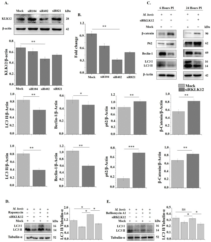
Knockdown of KLK12 impairs autophagy induction in M. bovis C68004 infected murine BMDMs. (A) Murine BMDMs were transfected with three different KLK12 siRNAs, having variable silencing targets, and a negative control siRNA (20 μM). After 48 h of transfection, KLK12 expression was determined by the Western Blot analysis. KLK12 expression was normalized to β-Actin expression; (B) murine BMDMs were transfected as stated above and after 48 h of transfection KLK12 expression was determined by qRT-PCR; (C) after 48 h of transfection, murine BMDMs were infected with M. bovis C68004 at a MOI of 10 and incubated for 6 or 24 h. Expression of autophagic markers LC3 II, Beclin-1, p62 and β-Catenin was determined in KLK12 siRNA and negative control siRNA transfected BMDMs by the Western Blot analysis. Expression of all the proteins was normalized to β-Actin expression; (D) murine macrophages were transfected as stated above and after 48 h of transfection, macrophages were treated with rapamycin (5 µm) for three h and then infected with M. bovis C68004 (MOI 10). After 24 h of infection, LC3 II expression was determined by WB; (E) murine macrophages were transfected as stated above and after 48 h, macrophages were treated with bafilomycin A1 (100 nm) for three h and then infected with M. bovis C68004 (MOI 10). After 24 h of infection, LC3 II expression was determined. Data represent the mean ± SD of three independent experiments (* p < 0.05; ** p < 0.01; *** p < 0.001).

