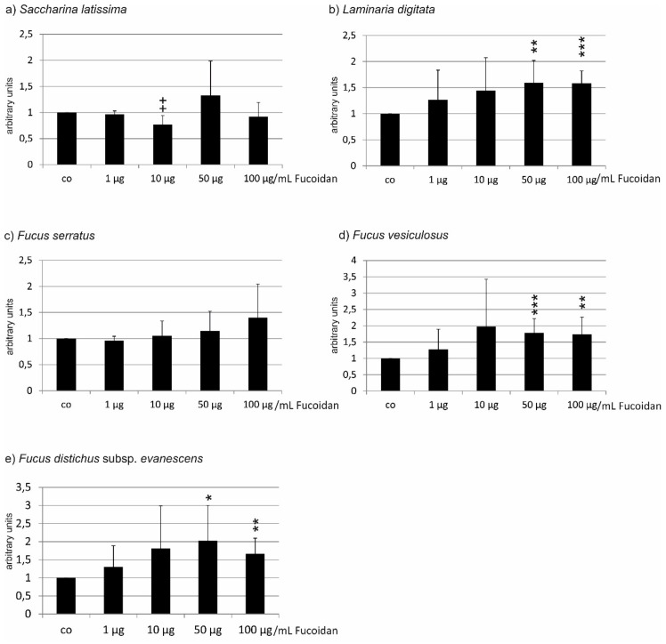
Figure 6.
VEGF secretion of primary porcine RPE cells after incubation with different concentrations of fucoidan from (a) Saccharina latissima (SL), (b) Laminaria digitata (LD), (c) Fucus serratus (FS), (d) Fucus vesiculosus (FV), (e) Fucus distichus subsp. evanescens (FE). VEGF content was evaluated in ELISA and normalized to cell viability. Control = 1. In RPE cells, only SL fucoidan reduced the VEGF content of RPE cells (10 µg/mL), while LD, FV, and FE induced a higher signal at concentrations of 50 and 100 µg/mL. Significance was evaluated with Friedman’s ANOVA and subsequent Student’s t-test, + p < 0.05 reduction compared to the control, * p < 0.05, ** p < 0.01 and *** p < 0.001 (n = 7).

