Figure 2.
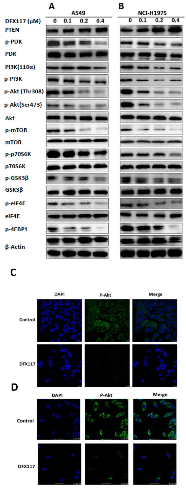
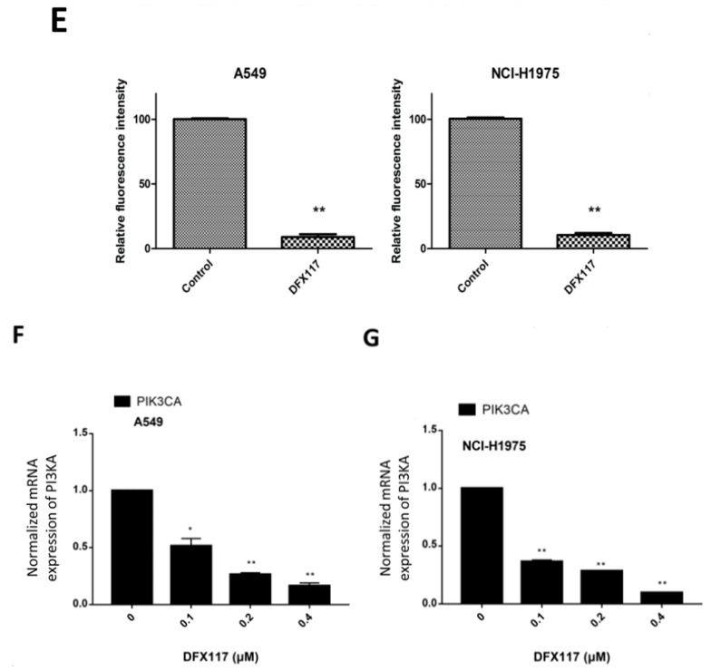
DFX117 suppresses the PI3K signaling pathway. (A,B) DFX117 suppressed the PI3K- signaling pathway in A549 and NCI-H1975 cells. Cells were collected for Western blot analysis after DFX117 treatment for 24 h at the indicated concentrations. The alterations in PI3K signaling related protein levels were quantified using Image J software. (C,D) Immunofluorescence analysis was performed to determine the effects of DFX117 on the expression level of p-Akt (Ser473) in A549 and NCI-H1975 cells. The cells were treated with DFX117 (0.2 μM) for 8 h, and then p-Akt expression was observed under a fluorescence microscope (magnification: 400×). DAPI staining was used to visualize and characterize the nucleus (blue). (E) Quantification of the fold changes in the expression levels of p-Akt is compared to the vehicle-treated control (lower panel). (F,G) DFX117 decreased the mRNA levels of PIK3CA in A549 and NCI-H1975 cells. Cells were treated with DFX117 for 24 h and then collected for RT-PCR analysis. * p < 0.05 or ** p < 0.01 was considered statistically significant compared with the corresponding control values.


