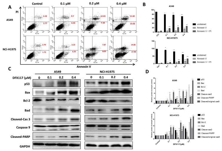
Figure 5.
DFX117 induced apoptosis in lung cancer cells. (A) Cell apoptosis was analyzed by flow cytometric analysis after Annexin V-FITC/PI staining. Cells were collected and centrifuged at 2000 rpm for 10 min after DFX117 treatment at various concentrations for 48 h. (B) The number in the right quadrant of each panel represents the percentage of Annexin V-positive cells. (C,D) The effect of DFX117 on the induction of apoptosis was also analyzed by Western blot in A549 and NCI-H1975 cells. The changes in corresponding protein expression levels were quantified using Image J. Each bar represents the mean ± SEM (n = 3). * p < 0.05 or ** p < 0.01 was considered statistically significant compared with the corresponding control values.

