Figure 6.
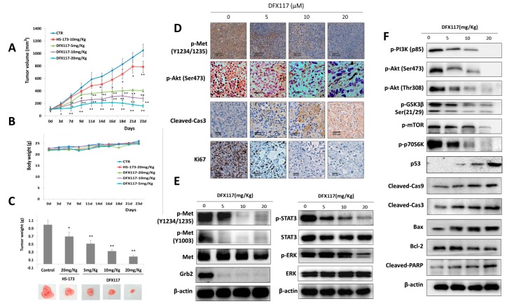
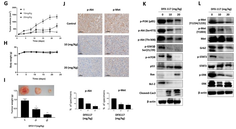
DFX117 inhibited tumor growth in a nude mouse xenograft model. (A–C) 3 × 10⁶ cells (A549) were subcutaneously injected into nude mice. Mice were orally administered DFX117 (5 mg/kg, 10 mg/kg and 20 mg/kg) or the vehicle control (DMSO: polyethylene glycol: saline = 1:5:4) daily for 23 days after tumor volumes reaching to 100 mm3 around. Tumor volumes were measured with calipers and body weights were monitored every 2–3 days. Data represent the means ± SEM (n = 5). (D) Determination of the alteration of p-Akt (Ser473), p-Met, Ki67 and cleaved caspase 3 in tumor tissues (A549 xenograft). The changes of the related proteins were evaluated by immunohistochemical analysis. Each tissue section was incubated with the indicated antibodies and analyzed by immunohistochemical method described in the Materials and Methods. (E,F) The effects of DFX117 on the expression levels of the corresponding proteins in Met/PI3K signaling in tumor tissues (A549 xenograft) were examined by Western blot analysis. (G–I) Mice bearing subcutaneously injected 7 × 10⁶ cells (NCI-H1975) were orally administered DFX117 (10 mg/kg or 20 mg/kg) or the vehicle control (DMSO: polyethylene glycol: saline = 1:5:4) daily for 23 days. Tumor volumes were measured with calipers, and body weights were monitored every 2–3 days. Data represent the means ± SEM (n = 5). (J) Immunohistochemical analysis of the expression level of p-Akt, p-Met in tumor tissues (NCI-H1975 xenograft). Each tissue section was incubated with the indicated antibodies and analyzed by immunohistochemical method described in the Materials and Methods. (K,L) The inhibition of DFX117 on Met/PI3K signaling in tumor tissues were analyzed by western blotting (NCI-H1975 xenograft). Data are representative of three independent experiments. Each bar represents the mean ± SEM (n = 3). * p < 0.05 or ** p < 0.01 was considered statistically significant compared with the corresponding control values.


